Introduction

Transrectal ultrasound-guided prostate biopsy (TRUS-Bx) is considered the current diagnostic standard for patients with suspected prostate cancer (PCa) based on raised serum prostatic specific antigen (PSA) levels, abnormal digital rectal examination findings and other risk factors.
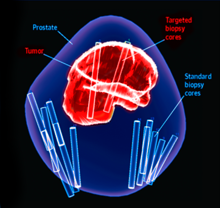
Recent years have seen significant advances in magnetic resonance imaging (MRI) technology. Increasing evidence suggests that MRI could non-invasively improve PCa visualization and aid in targeting prostate biopsies to abnormal areas seen on MRI. Hence, an approach with a pre-biopsy MRI followed by targeted biopsy (TBx), namely the “MRI pathway”, has become feasible, and studies have shown that MRI-targeted biopsy may improve detection of PCa.


Role of MRI in prostate cancer detection

Magnetic resonance imaging (MRI) is increasingly used as standard of care for pre-biopsy risk stratification in men with suspected prostate cancer due to its potential advantages over ultrasound.
For instance, in the recent PROMIS trial, multiparametric MRI combining T2-weighted imaging, diffusion-weighted imaging and dynamic contrast-enhanced MRI showed good sensitivity (88%) and negative predictive value (76%), and a meta-analysis on the updated Prostate Imaging Reporting and Data System (PI-RADS) version 2 showed sensitivity of up to 90% for detecting clinically significant prostate cancer.

The purpose of this page is to provide an interactive, up-to-date meta analysis and systematic review of prospective, randomized trials investigating the performance of MRI targeted biopsies for detecting PCa in patients compared to TRUS biopsies without MRI guidance.
The conclusion based on the overall data below is that prebiopsy MRI followed by MRI-guided biopsy will improve detection and risk stratification of PCa.

The latest data

Last updated 2019-03-18

Questions? Feedback?

Let us know what you think!